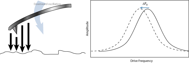
Electric Force Microscopy is analogous to standard Magnetic Force Microscopy (MFM) except that gradients being sensed are due to electrostatic rather than magnetic forces. In both methods, the cantilever is vibrated near its resonant frequency by a small piezoelectric element. The cantilever’s resonant frequency changes in response to any additional force gradient. Attractive forces make the cantilever effectively “softer,” reducing the cantilever resonant frequency. Conversely, repulsive forces make the cantilever effectively “stiffer,” increasing the resonant frequency:
Figure 1: Attractive gradient equivalent to additional spring in tension attached to the tip, reducing the cantilever's resonant frequency.
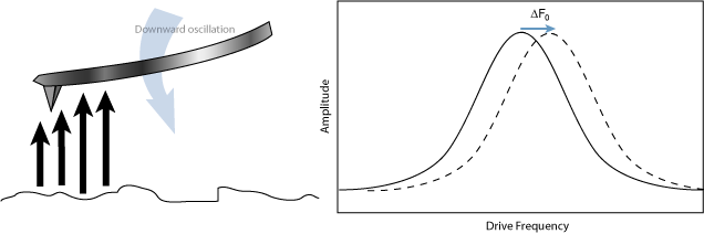
Figure 2: Repulsive gradient equivalent to additional spring in compression attached to the tip, increasing the cantilever's resonant frequency.
Changes in cantilever resonant frequency are detected in one of the following ways:
All of the above methods rely on the change in resonant frequency of the cantilever due to vertical force gradients from the sample. Figure 3 illustrates how the NanoScope V controller provides signal enhancement and feedback allowing gradient detection:
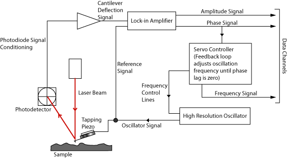
Figure 3: Block Diagram Of Controller Signals for EFM
The best candidates for electric field gradient imaging are samples that have large contrasts in the electric force gradient due to material differences or regions at substantially different potentials. For other samples having rough surface topography or small voltage variations this technique may be undesirable because topographic features will appear in the LiftMode image.
In many cases, you must apply a voltage to the tip or sample to achieve a high-quality image. Samples with permanent electric fields may not require voltage application.
| www.bruker.com | Bruker Corporation |
| www.brukerafmprobes.com | 112 Robin Hill Rd. |
| nanoscaleworld.bruker-axs.com/nanoscaleworld/ | Santa Barbara, CA 93117 |
| Customer Support: (800) 873-9750 | |
| Copyright 2010, 2011. All Rights Reserved. |
 Related Topics